The aim of brain tumour surgery is to remove the tumour tissue as completely as possible, but to spare areas of the brain that control sensation or movement, for example. With the help of an innovative imaging method, a team from the Department of Neurosurgery at the University Hospital Schleswig-Holstein (UKSH), Lübeck Campus, is working on distinguishing these functional areas and the tumour tissue even more precisely during the operation. This could further improve treatment outcomes for patients. The joint project "Intraoperative functional optical coherence tomography in neurosurgery combined with optical tumour localisation" combines the expertise of neurosurgery, the Medizinisches Laserzentrum Lübeck gGmbH and the Institute of Biomedical Optics at the University of Lübeck. German Cancer Aid is supporting the project over three years with a total of 581,000 euros as part of the "Surgery of the Future" funding initiative.
Optical coherence tomography (OCT) is an imaging technique that uses light waves to visualise the structure of tissue up to three millimetres deep in two- and three-dimensional images. It has proven to be a promising method for showing more precise boundaries between tumour and healthy tissue during brain tumour surgery. It also allows the high-resolution visualisation of vessels using Doppler measurements, similar to Doppler echocardiography, which can be used to visualise blood flows. Functional areas in the brain can be recognised, among other things, by the fact that they are supplied with more blood during activity, for example movement.
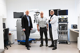
As part of the Neuro-OCT research project, funded by the German Federal Ministry of Education and Research and running since 2017, a so-called MHz (megahertz) OCT system, which enables several million depth scans per second, was integrated into a surgical microscope. The research group led by Dr Matteo Bonsanto, senior physician at the Clinic for Neurosurgery, Lübeck Campus, used the surgical microscope developed in Lübeck to carry out the world's first and only applications for intraoperative tumour detection in patients. This system is now being further developed as part of the project funded by Krebshilfe. Among other things, the project is investigating how real-time volume measurements of blood flow changes following electrical stimuli can improve the differentiation between functional areas, particularly for movement control, and tumour tissue.
"We are convinced that the use of the MHz-OCT system can sustainably improve safety during surgery and the quality of life of patients after surgery," says Dr Bonsanto, who is leading the project together with Prof. Dr Robert Huber from the Institute of Biomedical Optics and Dr Ralf Brinkmann from the Medical Laser Centre Lübeck.
The extent of tumour removal has a significant impact on the survival time of patients and the risk of tumour recurrence. However, extensive tumour removal near the motor centre, for example, also increases the risk of complications after the procedure, such as restricted movement and paralysis. Particularly in the case of gliomas, the most common brain tumours, and metastases, distinguishing between tumour tissue and healthy brain tissue during surgery is still a challenge. Technologies that are already used for this purpose, such as electrophysiology, fluorescent dyes and neuro-navigation, often reach their limits. False positive or negative signals in electrophysiological measurements or insufficient uptake of fluorescent dyes limit the accuracy of the results.
The Department of Neurosurgery, Lübeck Campus, is part of the University Cancer Centre Schleswig-Holstein (UCCSH), an association of all oncological facilities of the UKSH and the universities in Kiel and Lübeck.