Highlights & Galerie
Auszug aus den Forschungshighlights der Arbeitsgruppe
Seit der Gründung der Forschungsgruppe um Robert Huber im Jahr 2007 sind zahlreiche Meileinsteine in der Forschung um FDML-Laser, OCT-Bildung oder die nichtlineare Optik gesetzt worden. Darunter sind nicht nur forschungsbezogene Ereignisse sondern auch technologische Sprünge die hier ihre Erwähnung finden sollen.
Allgemein
James G. Fujimoto, Eric A. Swanson & Robert Huber - Hochauflösende medizinische Bildgebung
2017
Optische Kohärenztomografie
Erster Einsatz des Neuro-OCT-Systems im Operationssaal für die In-vivo-Bildgebung bei der Resektion von Hirntumoren
2024
Erste Veröffentlichung zum LARA-OCT Projekt für die großflächige OCT Aufnahme
2024
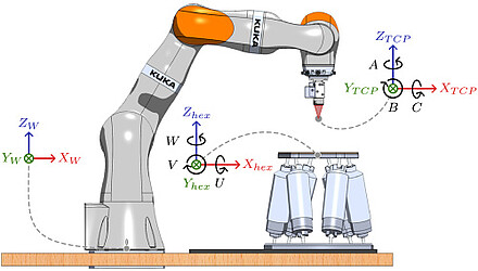
Wir demonstrieren die großflächige robotergestützte optische Kohärenztomographie (LARA-OCT), bei der ein Roboterarm mit sieben Freiheitsgraden in Verbindung mit einem 3,3-MHz-Swept-Source-OCT zur Rasterabtastung von Proben beliebiger Form verwendet wird. Wir haben eine Echtzeitkontrolle der Ausrichtung der Sonde zur Oberfläche implementiert, die die Sonde orthogonal zur Probe ausrichtet, indem sie nur Oberflächeninformationen aus den OCT-Bildern verwendet. Wir präsentieren OCT-Datensätze mit Volumengrößen von 140 × 170 × 20 mm3, die in 2,5 Minuten aufgenommen wurden.

Erste VR-OCT begleitete ex-vivo Operation an einem Schweineauge
2019
OCT video of human retina
2015
The video displays an OCT recording of a human retina. It was recorded at a volume rate of 20.8 V/s and consists of 255x255x450 voxels. Intentional movements of up to 100°/s are resolved.
High definition live 4D-OCT in vivo: Triops
2014
The video was acquired and processed in real time. The OCT system uses a 1310 nm Fourier domain mode locked (FDML) laser operated at 3.2 MHz sweep rate. The acquired data was processed and visualized on a GPU [10.1364/BOE.5.002963].
High definition live 4D-OCT in vivo for surgical guidance
2014
The video was acquired and processed in real time. The OCT system uses a 1310 nm Fourier domain mode locked (FDML) laser operated at 3.2 MHz sweep rate and the data was processed and visualized on a GPU [10.1364/BOE.5.002963].
3D reconstruction of intravascular OCT scan with 3200 fps
2013
A micromotor driven catheter developed in the group of Dr. Gijs van Soest in combination with our 1.6 MHz FDML-OCT surpasses the acquisition speed of state of the art intravascular OCT by more than one order of magnitude [10.1364/OL.38.001715].
Anterior segment imaging with extended coherence length MHz FDML laser
2012
OCT imaging at 60 nm sweep range and 1.6 MHz scan rate. The 3D reconstruction of the whole anterior segment consisting of 1000 x 985 x 560 voxels (frames x depth scans x samples/scan) was acquired in a total time of 0.8 seconds [10.1364/BOE.3.002647].
Anterior segment imaging with extended coherence length MHz FDML laser
2012
The 3D reconstruction of the whole anterior segment consisting of 1000 x 900 x 560 voxels (frames x depth scans x samples/scan) was acquired in a total time of 0.8 seconds [10.1364/BOE.3.002647].
High quality 3D imaging at 20 million A-scans and 4.5 GVoxels per second
2010
This three dimensional dataset was acquired in 25ms. This was accomplished by using a FDML laser with a sweep rate of 5.2 MHz per spot and a 4-spot illumination [10.1364/OE.18.014685].


